What We Can Learn From Their X-Rays About Our Teeth
We will likely take a variety of dental x-rays during your or your child's initial visit to our Summerlin location. Thanks to these films, we can view the inner structure of your mouth, including the bone, the roots of your teeth, and the nerve tissue that connects them.
Radiographs are incredibly useful for diagnosing dental conditions that are not visible to the naked eye. These disorders can affect the teeth and jaw. For instance, if you are experiencing pain but there are no visible abnormalities in your mouth, dental x-rays will reveal whether or not an infection is growing beneath the surface of the tooth or teeth causing the suffering.
We utilize four different types of x-rays, including bitewings, periapicals (PAs), a panorex or panoramic, and a dental cat-scan. Due to the specific characteristics of each form of dental radiograph, we are able to accurately diagnose an extensive range of dental problems. Following is a breakdown of the elements that comprise each film, as well as an explanation of why we want a specific image.
When you brush your teeth, the majority of the plaque and tartar buildup on your smile will be eliminated. However, the bristles cannot reach the spaces between your teeth to remove plaque from those regions. Therefore, the dentist at our Summerlin office strongly advises patients to floss at least once every day. In the case that they are not removed, the leftover food particles will begin to erode your enamel, necessitating a dental procedure to remove it.
We rely on bitewing x-rays to offer light on this aspect of your evaluation since we cannot detect what is occurring between your teeth by simply observing your mouth. Once a year, at one of your twice-yearly dental cleanings, we will take this series of radiographs as a matter of routine and policy.
Taking these images is a quick and straightforward process. When it comes to small children, we can collect all the necessary information from two x-rays. When photographing adults, four photographs are taken. This particular radiograph depicts the body of the tooth structure, but there is no trace of anything below the gumline.
Periapical Films
If we have reason to assume that your dental problem is located deeper in your tooth, the periapical x-ray will reveal the whole anatomy of the tooth, including the pulp and nerve canals, as well as the surrounding roots and bone. In this instance, the periapical x-ray will be taken.
If we are dealing with a tooth with an abscess, this film will display a black shadow at the end of the root. In addition to aiding in the diagnosis of your condition, a periapical examination will provide the information necessary to identify the most efficient approach of treatment. For example, if there is adequate healthy tooth structure remaining and the nerve anatomy within your tooth is typical, we will likely recommend root canal therapy to fix it. This will enable us to eliminate the infection and preserve your tooth. If the x-rays taken at the dentist's office reveal that too much of the tooth is gone, the tooth will need to be pulled.
When we inspect the tooth's roots, it will be easier for us to determine whether or not you need to see an oral surgeon to have a tooth out. If the roots are very large and curved, or if there are significant nerves flowing through the area, you may be referred to a specialist for your own protection.
A Panorex ("Pano")
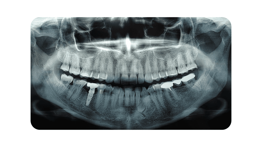
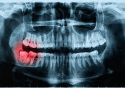
When your child reaches a certain age, we will verify whether their teeth are developing regularly and in the proper sequence. In order to aid in this evaluation, we will take a panorex x-ray, which is a two-dimensional image that displays the head and neck's inner architecture from the nose to the chin and all the way back to the TMJ.
In addition, wisdom teeth, which might be difficult to detect with other forms of x-rays, are far easier to inspect with panorex images. In addition, these photographs will reveal any bone fractures, malignancies, sinus infections, or cysts that may be present.
Dental Cat-Scan

A dental CT scan delivers the same information as a panorex, but because it generates a three-dimensional image, it provides far more detail. This image can be used to establish the amount of space in the bone, the density of the bone, and whether dental implants are a realistic option for you. If you are considering dental implants, this image can be used. During the implant placement process, the CT scan is crucial in ensuring that the implant is accurately positioned.
Because this three-dimensional imaging allows us to see the exact shape of the tooth's nerves and roots, it is extremely useful for root canal therapy and extractions. As a direct result of this, root canal treatments have a better success rate, and tooth extractions are easier to do.
Trust Our Smile Team
Dr. Cohan, our Summerlin dentist, uses the latest advanced digital x-ray technology to precisely detect and treat any dental diseases his patients may have. When you become a patient of ours, the care of your health will be our top priority. By integrating cutting-edge technology, we can ensure that we are doing everything possible to improve your smile and general health.
Residents of the Summerlin region who have been searching for a new dentist should visit our office immediately. Call us immediately to schedule an appointment!
CLICK HERE TO SCHEDULE YOUR APPOINTMENT!
GET TO KNOW YOUR BEST DENTIST IN SUMMERLIN
Dr. Marianne Cohan was voted The Best Dentist/ Dental Office and Best Cosmetic Dentist from The Las Vegas Review-Journal in 2020 and 2021. She received her Doctor of Dental Surgery (DDS) from the State University of New York at Buffalo in 1992.

With an emphasis on cosmetic dentistry, complete makeovers, and implant dentistry, Dr. Cohan is committed to continuing education and feels that we never stop learning. Dr. Cohan takes pride in using high-powered magnification to perform minimally invasive restorative dentistry. She uses all the latest technological advances including digital radiography, digital photography, computer simulations, and high-resolution pictures of your proposed treatment on 55-inch screens. She also utilizes CBCT (cone beam) and laser technology.
Dr. Cohan is always available to her patients and is available for any dental emergency.